Surgical and medical

Skin cancer, growths and spots that need a diagnosis, treated with a surgeon's precision and a cosmetic eye. Dr. Nawas is a fellowship-trained Mohs surgeon, and Original Medicare is accepted for medical and surgical care.

How it works here

From first look to treatment.

  1. Skin check.

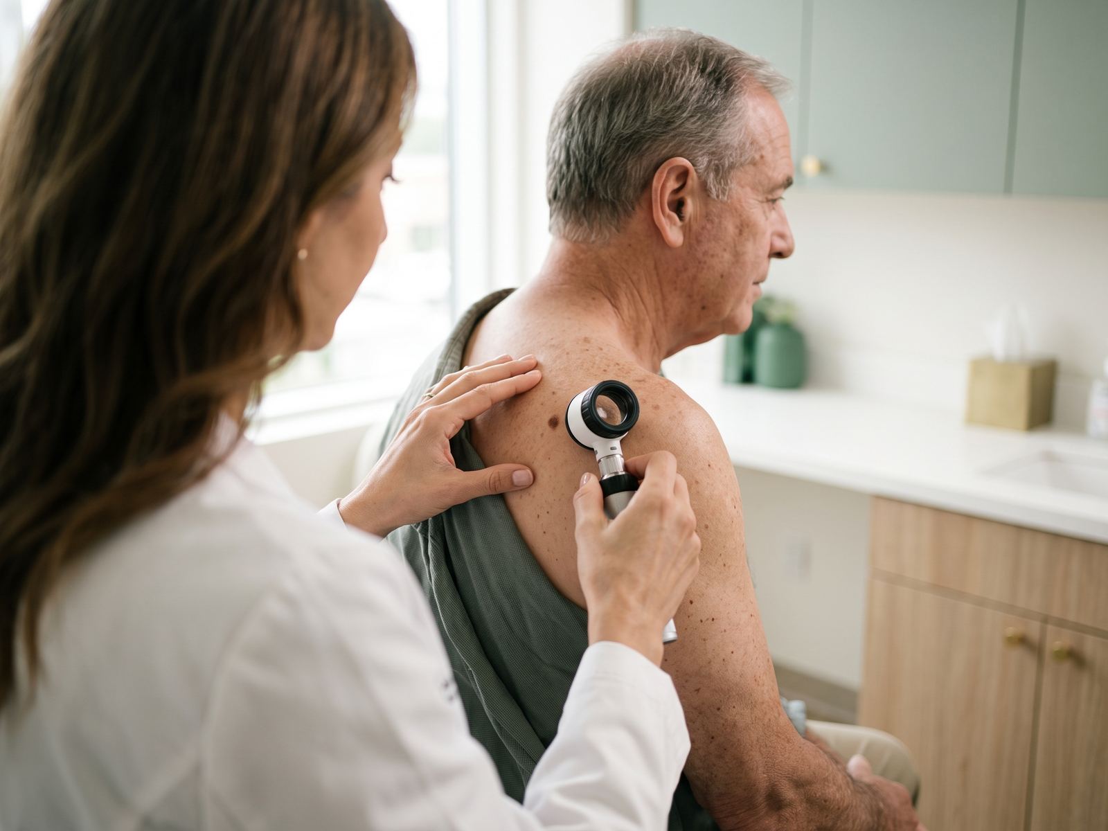
    A full skin exam and skin cancer screening with Dr. Nawas, who looks at every spot that concerns you.

  2. Biopsy, when a spot needs a diagnosis.

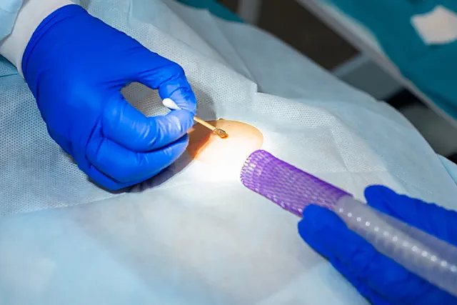
    A tiny sample, taken in minutes, so the diagnosis rests on evidence rather than appearance. Results usually take about a week. Read about skin biopsy.

  3. Treatment matched to the diagnosis.

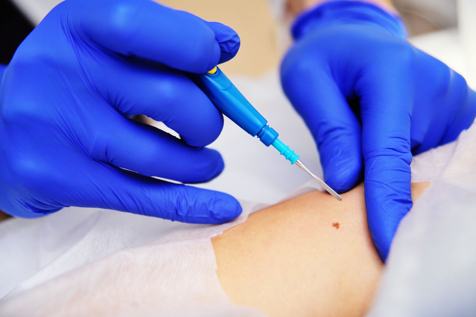
    Mohs micrographic surgery, Surgical excision, Cryotherapy, Electrodesiccation.

The treatments

What each one does.

Skin cancer surgery

Mohs micrographic surgery

By a fellowship-trained Mohs surgeon

The most precise method for removing skin cancer, with the highest cure rates.

Treats
Basal cell and squamous cell carcinoma, especially on the face
Downtime
Stitches for 1 to 2 weeks
Pricing
Original Medicare accepted
Read the full guide

Growths and skin cancers

Surgical excision

Complete removal of a lesion with a repair planned for how it will look in a year.

Treats
Skin cancers on the body, moles, cysts, lipomas, torn earlobes
Downtime
Stitches for 1 to 2 weeks
Typical cost
$500 to $2,500 by procedure
Read the full guide

Diagnosis

Skin biopsy

Shave, punch or excisional biopsy

A small sample taken so a diagnosis is made on evidence rather than appearance.

Treats
Changing moles, unclear growths, rashes that will not settle
Downtime
A small dressing for several days
Typical cost
$300
Read the full guide

Precancers, warts and growths

Cryotherapy

Liquid nitrogen for precancerous and benign lesions

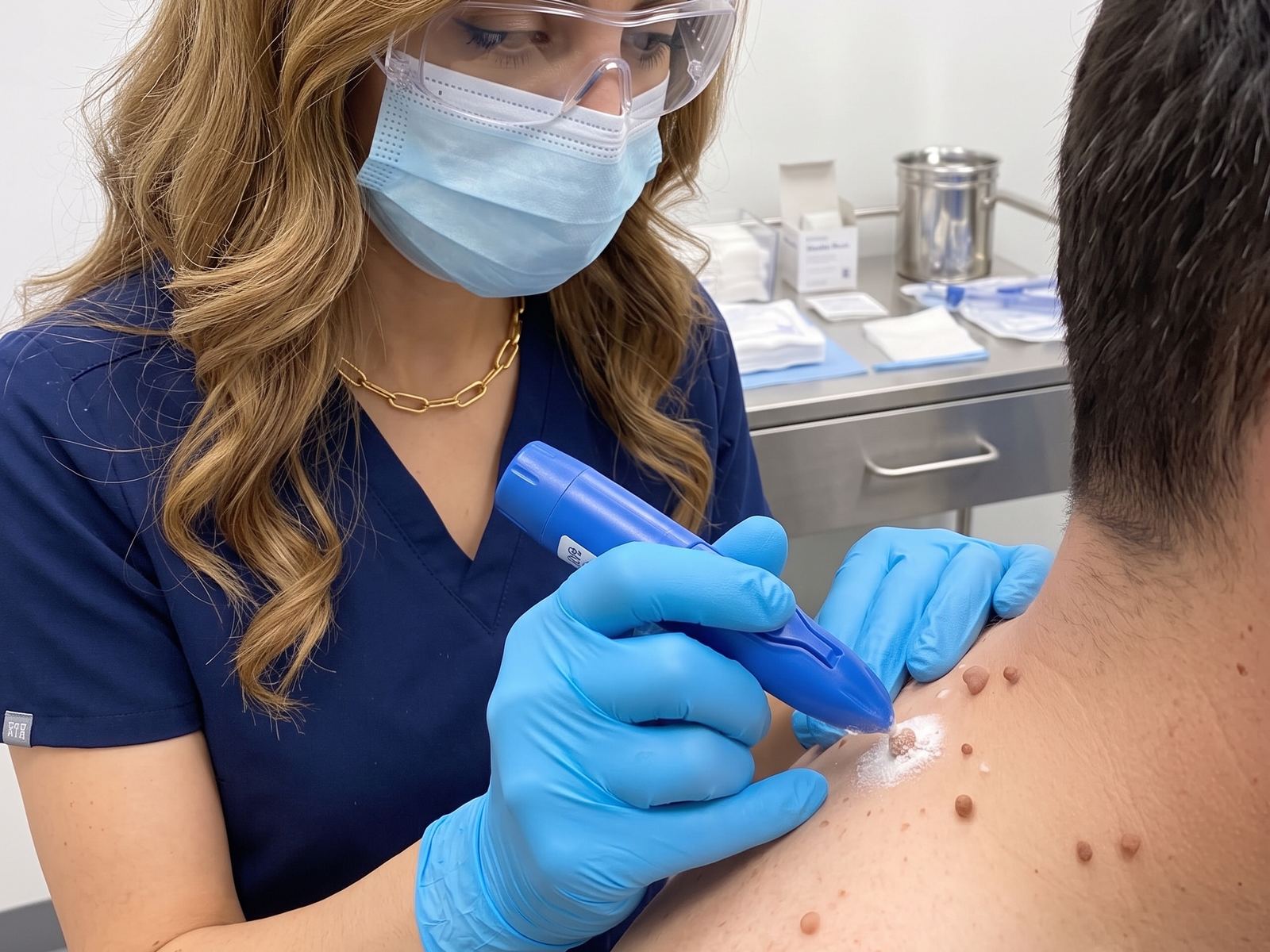
Liquid nitrogen used to destroy precancerous and benign lesions in seconds.

Treats
Actinic keratoses, warts, seborrheic keratoses, skin tags
Downtime
A blister or scab for 1 to 2 weeks
Typical cost
$200 per session
Read the full guide

Small harmless growths

Electrodesiccation

Small harmless growths dried and sealed with a fine electric needle, for skin that heals flat.

Treats
Skin tags, cherry spots, sebaceous hyperplasia, seborrheic keratoses, DPN
Downtime
Small crusts for 1 to 2 weeks
Typical cost
Up to $1,000
Read the full guide

How they work

Still from the animation: a cross-section of skin with a shave biopsy removing the raised top of a mole with a fine blade
How a skin biopsy works One minute, no sound
Read the steps

Why a biopsy. When a spot looks unusual, the only way to know what it is for certain is to look at its cells under the microscope. A biopsy takes a tiny sample to do exactly that.

  1. Numb. The area is numbed with a small injection of local anesthetic. After that, the biopsy itself is painless and takes only a few minutes.
  2. Sample. Depending on the spot, a shave biopsy removes the raised top with a fine blade, or a punch biopsy takes a small round core, usually closed with one or two stitches.
  3. Examine. The sample goes to a dermatopathology lab, where it is sliced thinly, stained and examined under the microscope. Results usually take about a week.
  4. Heal and plan. The site heals in one to two weeks, leaving a small mark that fades. The result guides what, if anything, comes next.
Still from the animation: a cross-section of skin with the first thin layer removed, the tissue on its way to the lab for frozen sections, and the color-coded Mohs map of its edges
How Mohs surgery works One minute, no sound
Read the steps

Why Mohs. Skin cancers often send out roots that cannot be seen from the surface. Mohs surgery removes the cancer in thin layers and checks every edge under the microscope, the same day.

  1. Remove a layer. After numbing, the visible cancer is removed with a thin rim of normal-looking skin. The piece is mapped and color-coded so each edge can be traced back to its exact spot.
  2. Check every edge. While you wait, the tissue is frozen, sliced and stained in the lab, then examined under the microscope. The entire edge and underside are checked, not just samples.
  3. Repeat only where needed. If cancer remains at one point on the map, only that spot is removed in the next layer. This continues until the edges are clear, taking the least healthy skin possible.
  4. Repair. Once the margins are clear, the wound is repaired the same day with the technique that best preserves function and appearance. Cure rates for common skin cancers are among the highest of any treatment.
Still from the animation: a cross-section of skin with a scalpel removing a growth in one piece, with a thin margin of normal skin around it
How a skin excision works One minute, no sound
Read the steps

When it is used. An excision removes a growth completely, with a thin margin of normal skin around it: a cyst, an irregular mole, or a skin cancer that is best treated with a clear margin.

  1. Plan and numb. The area is cleaned and the shape is marked, usually a narrow ellipse along the natural skin lines so the scar settles into a crease. Local anesthetic makes the procedure painless.
  2. Remove. The marked skin is removed in one piece, down to the fat, and sent to the lab to confirm the diagnosis and that the edges are clear.
  3. Close. The wound is closed in layers: dissolving stitches beneath the surface take the tension, and fine stitches on top line up the edges precisely.
  4. Heal. Surface stitches come out in about one to two weeks. The scar is pink at first and fades over 6 to 12 months; silicone gel and sun protection help it along.
Still from the animation: a cross-section of skin with a growth frozen white by liquid nitrogen and starting to thaw
How cryotherapy works One minute, no sound
Read the steps

What it treats. Cryotherapy uses liquid nitrogen, colder than 300 degrees below zero Fahrenheit, to treat warts, precancerous actinic keratoses, seborrheic keratoses and other small growths.

  1. Freeze. A spray of liquid nitrogen is aimed at the growth for a few seconds. Ice forms inside the abnormal cells and breaks them apart, while the surrounding skin is spared.
  2. Thaw. The frozen spot turns white, then thaws over a minute or so. It stings during the freeze and thaw, then settles. Some lesions are frozen twice in the same visit.
  3. Blister and crust. Over the next day or two the area may swell or blister, then forms a crust. The crust separates on its own within one to two weeks.
  4. Heal. Fresh skin forms beneath the crust. The treated spot may be lighter or pinker for a while, and thicker growths sometimes need a second session.
Still from the animation: a cross-section of skin with a fine needle tip delivering a tiny current to a small raised growth, drying and sealing it
How electrodesiccation removes growths One minute, no sound
Read the steps

What it treats. Electrodesiccation is used for small benign growths that sit in the top layers of the skin: sebaceous hyperplasia, skin tags, seborrheic keratoses and similar spots.

  1. Numb. The spot is numbed with a drop of local anesthetic, so the treatment itself is comfortable and takes only seconds per lesion.
  2. Dry. A fine needle tip delivers a tiny electrical current to the growth. The current dries and seals the tissue precisely, without touching the healthy skin around it.
  3. Crust. The treated growth becomes a small dry crust. It separates on its own over the following week or two; picking it is the one thing to avoid.
  4. Heal. Underneath, fresh skin has already formed. The spot heals flat and smooth, and any pinkness fades over the following weeks.